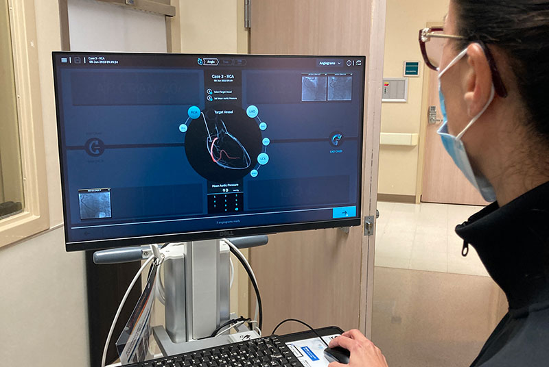
White Rock Medical Center is the first in Texas to use artificial intelligence-based technology for care of patients with heart disease.
The AI-based system helps physicians safely and precisely view and calculate a heart blockage so they can determine how to proceed in as little as 45 seconds while bedside in the cardiac catheterization lab.
Called CathWorks FFRangioTM, the new system improves upon an already existing test called FFR. Fractional flow reserve (FFR) measures the pressure and flow of blood in coronary arteries to help determine the location and degree of blockage.
The process takes several steps, but in reality, the 3D image and diagnostic information are ready in less than a minute.
“The FFRangio system is a new tool to gather objective data to ensure the patient receives the appropriate level of care and treatment, such as the diameter and the length of the stent,” explained Mahmood Ali, MD, a cardiologist who worked on the first FFRangio. “Angiography is a great way to look at the vessels but it’s not without limitations. What sets this new FFRangio system apart is its speed and ease of use while obtaining additional information,” he said.
Typically, a minimally invasive FFR test involves inserting a wire into an artery. The procedure can help the healthcare provider determine degree of blockage. In turn, the information is used to make decisions about the patient’s medical treatment.
The new system at White Rock Medical Center is unique in that it doesn’t require use of a wire, produces real-time three-dimensional (3D) images and calculates blood flow and blockage.
After an angiogram produces 2D, X-ray images of the blood vessels, those images are loaded into the new FFRangio system. The provider reviews the 2D images and marks the targeted area of blockage. The AI-based system traces the vessels and the area of lesion. It then converts the images to a 3D rendering, matching vessels and location of the lesion across the various 2D images. The system then produces an FFR calculation and color-coded, 3D image of the artery.
“An FFR without a wire in the artery lowers risks and complications. This is comparatively less invasive and still determines flow restrictions,” said Ron Bullard, manager of White Rock Medical Center’s cardiac cath lab. “What it produces is diagnostic information that can then be used to determine a course of action for the patient’s care.
“We’re the only healthcare center doing 3D, multivessel angio-based images in Texas,” added Bullard.
White Rock Medical Center holds a Certificate of Distinction for Chest Pain as well as Primary Stroke Center Advanced Certification by The Joint Commission.

